BioAcyl Corp |
|
Brown, L. S., Foster, C. G., Courtney, J.-M., & others. (2019). Pericytes and neurovascular function in the healthy and diseased brain. Front. Cell. Neurosci. 13. Added by: Dr. Enrique Feoli (26/06/2021, 13:19) Last edited by: Dr. Enrique Feoli (26/06/2021, 13:30) |
| Resource type: Journal Article Published DOI: 10.3389/fncel.2019.00282 ID no. (ISBN etc.): 1662-5102 BibTeX citation key: Brown2019 View all bibliographic details |
Categories: BioAcyl Corp Subcategories: Neurovascular function Creators: Brown, Courtney, Foster, others Collection: Front. Cell. Neurosci. |
Views: 9/270
|
| Abstract |
|
Pericytes are multi-functional cells embedded within the walls of capillaries throughout the body, including the brain. Pericytes were first identified in the 1870s, but little attention was paid to them during the following century. More recently, numerous vascular functions of pericytes have been identified including regulation of cerebral blood flow, maintenance of the blood-brain barrier, and control of vascular development and angiogenesis. Pericytes can also facilitate neuroinflammatory processes and possess stem cell-like properties. Pericytes form part of the neurovascular unit, a collection of cells that control interactions between neurons and the cerebral vasculature to meet the energy demands of the brain. Pericyte structure, expression profile, and function in the brain differ depending on their location along the vascular bed. Until recently, it has been difficult to accurately define the sub-types of pericytes, or to specifically target pericytes with pharmaceutical agents, but emerging techniques both in vitro and in vivo will improve investigation of pericytes and allow for the identification of their possible roles in diseases. Pericyte dysfunction is increasingly recognized as a contributor to the progression of vascular diseases such as stroke and neurodegenerative diseases such as Alzheimer{'}s disease. The therapeutic potential of pericytes to repair cerebral blood vessels and promote angiogenesis due to their ability to behave like stem cells has recently been brought to light. Here, we review the history of pericyte research, the present techniques used to study pericytes in the brain, and current research advancements to characterize and therapeutically target pericytes in the future.
|
| Notes |
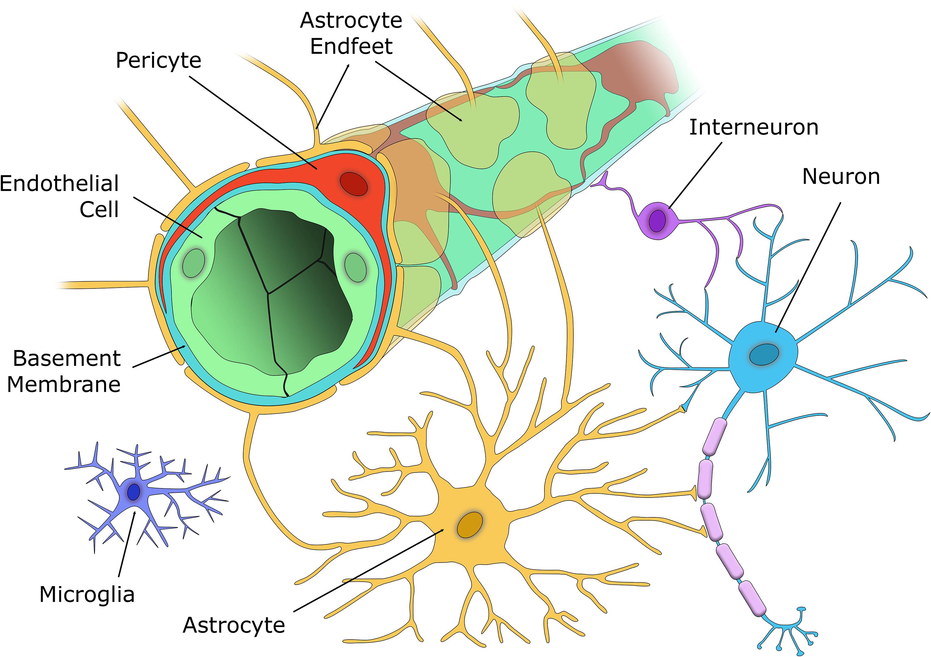
The cellular components of the NVU. The capillary wall contains a single EC layer (green) connected by tight junctions that form the blood-brain barrier along with pericytes (red), that are embedded within the capillary basement membrane (light blue), and nearby astrocyte endfeet (yellow). Excitatory neurons (blue) synapse with both vasoactive interneurons (purple), and astrocytes (yellow), who in turn signal to the capillary to alter blood flow according to the metabolic demands of that brain region. Microglia (indigo) are located in the brain parenchyma and respond to any aversive stimuli to protect the brain. Added by: Dr. Enrique Feoli Last edited by: Dr. Enrique Feoli |