BioAcyl Corp |
|
Fiore, A. P. Z. P., Ribeiro, P. D. F., & Bruni-Cardoso, A. (2018). Sleeping Beauty and the Microenvironment Enchantment: Microenvironmental Regulation of the Proliferation-Quiescence Decision in Normal Tissues and in Cancer Development. Frontiers in Cell and Developmental Biology, 6. Added by: Dr. Enrique Feoli (28/08/2022, 20:06) Last edited by: Dr. Enrique Feoli (14/01/2023, 11:36) |
| Resource type: Journal Article ID no. (ISBN etc.): 2296-634X BibTeX citation key: Fiore2018 View all bibliographic details |
Categories: BioAcyl Corp Subcategories: Microenvironment Creators: Bruni-Cardoso, Fiore, Ribeiro Collection: Frontiers in Cell and Developmental Biology |
Views: 2/217
|
| Abstract |
|
Cells from prokaryota to the more complex metazoans cease proliferating at some point in their lives and enter a reversible, proliferative-dormant state termed quiescence. The appearance of quiescence in the course of evolution was essential to the acquisition of multicellular specialization and compartmentalization and is also a central aspect of tissue function and homeostasis. But what makes a cell cease proliferating even in the presence of nutrients, growth factors, and mitogens? And what makes some cells “wake up” when they should not, as is the case in cancer? Here, we summarize and discuss evidence showing how microenvironmental cues such as those originating from metabolism, extracellular matrix (ECM) composition and arrangement, neighboring cells and tissue architecture control the cellular proliferation-quiescence decision, and how this complex regulation is corrupted in cancer.
|
| Notes |
|
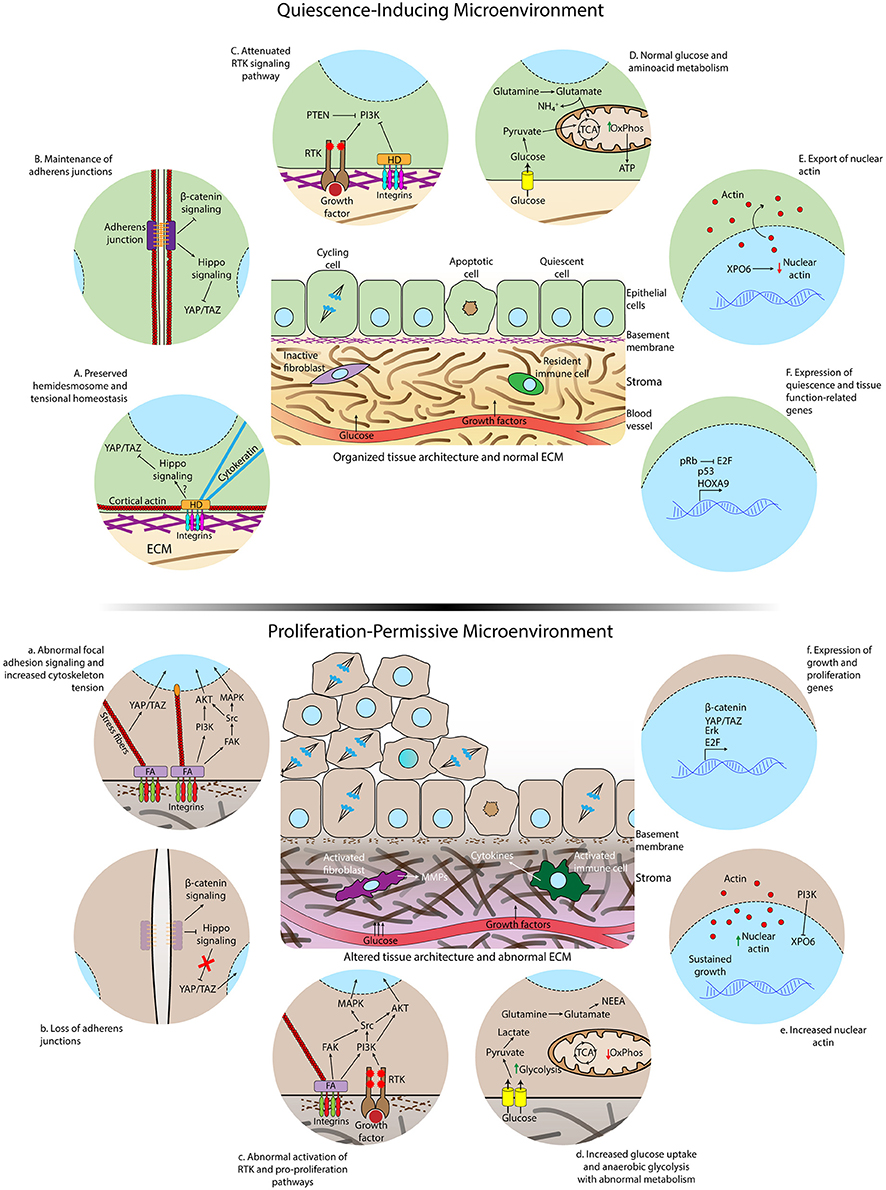
The Composition and Physical Properties of the Extracellular Matrix Are Key Determinants of Cellular Quiesce
Differences in microenvironmental and subcellular signaling between homeostatic conditions and loss of cell quiescence. (TOP) Quiescence-inducing microenvironment showing normal extracellular matrix, intact basement membrane, organized tissue architecture, and resident immune cells and fibroblasts. The rates of proliferation and apoptosis are comparable to maintain homeostasis. (BELOW) Altered and proliferation-permissive microenvironment displaying abnormal extracellular matrix with altered composition and structure, disrupted basal lamina, activated fibroblasts in the stroma, inflammatory infiltrate by activated macrophages and other cytokines-secreting activated immune cells. The rate of proliferation is increased due to loss of quiescence regulation by the tissue microenvironment. Details of epithelial cells residing in a healthy (A-F) or aberrant microenvironment (A–F). A normal ECM and correct tissue architecture induces the formation of hemidesmosomes connecting the ECM to cytokeratin filaments, cell-cell junctions, cortical actin cytoskeleton, and polarized epithelium (A,B). Consequently, the Hippo pathway, that inhibits translocation of YAP/TAZ to the nucleus, is activated, receptor tyrosine kinase (RTK) activity is attenuated (C) and nuclear actin export is enhanced (E). Glucose and glutamine are completely metabolized by the tricarboxylic acid cycle (TCA) and oxidative phosphorylation (OxPhos) (D). Quiescence gene expression programs are triggered during quiescence acquisition (F). Aberrant ECM signaling due to altered ECM composition and stiffness increases formation of focal adhesions (FA), loss of cortical distribution of the actin cytoskeleton, enhanced activation of FAK, formation of actomyosin stress fibers, translocation of YAP to the nucleus (A) and accumulation of nuclear actin (E). Loss of adherens junctions allows the translocation of beta–catenin to the nucleus (B). Overactivation of growth factor signaling occurs as a consequence of intersection between integrin and RTK-triggered signaling (C). Glucose uptake and anaerobic glycolysis are further increased due to exacerbated activation growth factor pathways, glutamine is converted to NEAA and biosynthetic precursors in the TCA and oxidative phosphorylation is reduced in the aberrant microenvironment (D). Activation of genes involved in cell-cycle entry is increased (F). |