BioAcyl Corp |
|
Silva-Sanchez, A., & Randall, T. D. (2020). Anatomical Uniqueness of the Mucosal Immune System (GALT, NALT, iBALT) for the Induction and Regulation of Mucosal Immunity and Tolerance. Mucosal Vaccines, 21. Added by: Dr. Enrique Feoli (29/09/2020, 18:41) Last edited by: Dr. Enrique Feoli (08/01/2023, 14:07) |
| Resource type: Journal Article Published DOI: 10.1016/B978-0-12-811924-2.00002-X BibTeX citation key: SilvaSanchez2020 View all bibliographic details |
Categories: BioAcyl Corp, BioAcyl Corp Subcategories: Inmunidad de mucosas, Sec lymphoid organs Creators: Randall, Silva-Sanchez Collection: Mucosal Vaccines |
Views: 5/414
|
|
Conclusiones
requires mechanisms for these cells to find each other and additional mechanisms to support their proliferation and survival during immune responses [14].
Added by: Dr. Enrique Feoli |
| Abstract |
|
Mucosal surfaces lining the lung and the gut are constantly exposed to environmental antigens, commensal organisms, and pathogens. Consequently, the host’s immune system devotes enormous resources to the defense of these tissues. Although mucosal immune responses must be sufficiently strong to eliminate pathogens, they must also have mechanisms to prevent excessive inflammation and initiate epithelial repair. Importantly, mucosal immune responses are initiated and regulated by a variety of mucosal lymphoid tissues, collectively known as mucosa-associated lymphoid tissue (MALT). This chapter describes the unique developmental, architectural, and functional features of the MALT and how those features facilitate the local immune response at each site.
|
| Notes |
|
Why is lymphoid organization important? The evolution of specialized antigen-presenting cells, B cells and T cells, all of which need to interact for successful immune responses, requires mechanisms for these cells to find each other and additional mechanisms to support their proliferation and survival during immune responses [14]. In this regard, the development of specialized stromal cells, including fibroblastic reticular cells (FRCs) in the T cell area and follicular dendritic cells (FDCs) in the B cell area, is likely a key innovation. These cell types provide the scaffolding of lymphoid organs and help to spatially organize lymphocytes and antigen-presenting cells. For example, FRCs express the chemokine CCL19, which attracts T cells and activated dendritic cells (DCs) into the T cell zone [18], [19], whereas FDCs express the chemokines CXCL12 and CXCL13, which attract B cells and T follicular helper (Tfh) cells and direct their spatial positioning in the B cell follicle and germinal center [20], [21], [22], [23]. Although B and T cell separation occurs in amphibians and reptiles, identifiable stromal cells such as FDCs are observed only in birds and mammals [14] (Fig. 2.1), which probably explains why these species (and not others) are able to support germinal center responses and generate high-affinity antibodies [24], [25]. Thus the mechanisms that control the differentiation of stromal cell elements are critical for the proper organization and function of lymphoid organs.
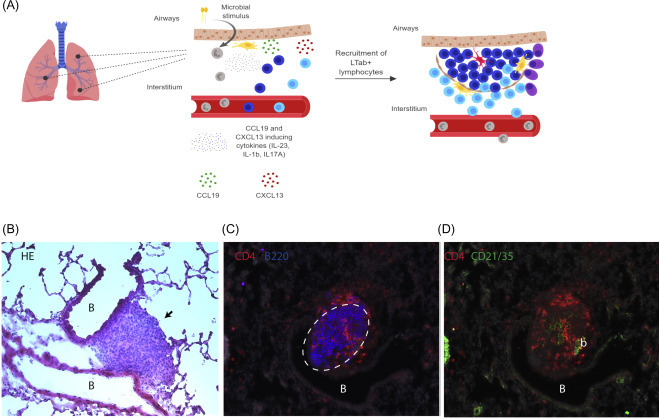
The common and distinct mechanisms of protective and pathogenic iBALT formation. 1) Protective iBALT is induced in response to PAMPs recognized by PRR expressed on innate immune cells or epithelial cells. Pathogenic iBALT is induced in response to chronic pathogen exposure, self-antigens, or environmental Ags, which induce DAMPs and inflammation. 2) The sensing of PAMPs and DAMPs results in the production of iBALT-driving cytokines that induce the local response of myeloid cells, production of inflammatory molecules, and recruitment of more myeloid cells, lymphocytes, FDCs, and Tfh. 3) These signals and associated cellular recruitment drive the formation and organization of GC. 4) In protective iBALT, responses are associated with early recruitment of neutrophils and cDCs, IFN-γ–mediated pathogen clearance, and short-term inflammation. In pathogenic iBALT, responses are associated with autoantibodies and uncontrolled inflammation, with some diseases specifically highlighted by eosinophilia-induced inflammation. Other changes include altered metabolic state and dysregulated lymphocyte proliferation with subsequent long-term unresolved inflammation. |
| Quotes |
|
Added by: Dr. Enrique Feoli
(30/09/2020, 16:06)
|
| Paraphrases |
What is an HEV?Lymphocytes enter lymph nodes directly from the blood by migrating across the walls of specialized postcapillary venules which are located in the paracortex (T lymphocyte areas) of the node. These vessels have a characteristic morphology which distinguishes them from other types of blood vessel. The endothelial cells which line these vessels have a plump or cuboidal morphology, which contrasts to the flattened morphology of endothelial cells lining other types of vessel (Figure 1). It is the peculiar endothelial morphology that has engendered their common name – high endothelial venules (HEVs). The endothelial lining is surrounded by a thickened basement membrane, which is more pronounced than in other types of blood vessel. In immunocompetent adults, the localization of lymphocytes, but not other leukocytes, either attached to the inner (luminal) surface or within the vessel wall and the distinct morphological appearance are the most recognizable features of HEV. However, these vessels are also readily distinguishable from other blood vessels by histochemical, biochemical and immunological markers. The lining endothelial cells, which will be referred to as high endothelial cells (HECs), stain more intensely with RNA-binding dyes such as pyronin and contain a prominent Golgi apparatus and more secretory vesicles than other types of endothelia. Early histochemical studies also distinguished HEC from ‘flat’ endothelial cells lining other types of vessel. HEC express higher levels of nonspecific esterase, lactate dehydrogenase and NADH reductase. Biochemical studies identified a unique biosynthetic pathway for inorganic sulfate in HECs which is crucial for the generation of the peripheral addressin, an important adhesion molecule which mediates l-selectin-dependent binding of lymphocytes to HECs (see below). Several monoclonal antibodies have been described that specifically stain HEVs and not other types of blood vessel. These include MECA 79 and MECA 367, which identify the peripheral and mucosal addressins respectively, HECA 452, which identifies part of the functional carbohydrate epitope in the peripheral addressin, the sialyl Lewisx saccharide and MECA 325, for which the antigen has not yet been identified. 
Added by: Dr. Enrique Feoli
(30/09/2020, 14:55)
|